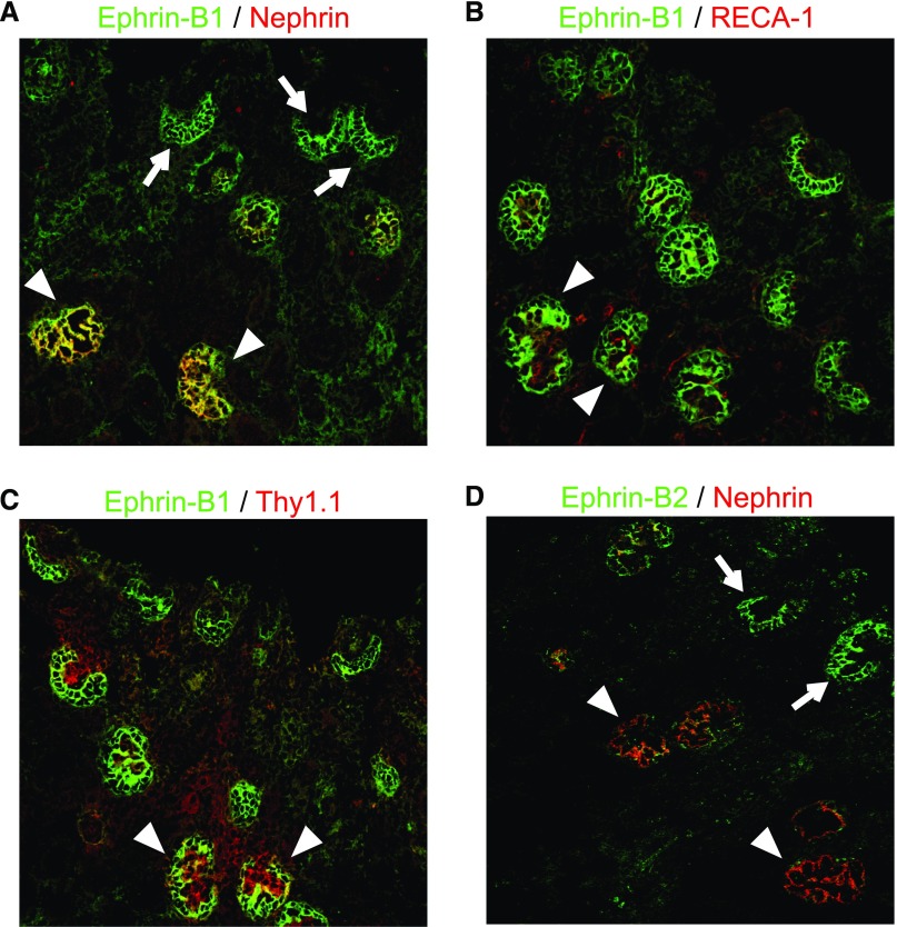
Figure 2.
Ephrin-B1 appeared in presumptive podocytes earlier than nephrin. Immunofluorescence findings with anti–ephrin-B1 (A–C) and anti–ephrin-B2 (D) antibodies (shown in green) costained with an anti-nephrin antibody, anti-RECA1 (endothelial cell marker) and anti-Thy1.1 (mesangial cell marker) (shown in red). (A) The staining of ephrin-B1 was first detected in presumptive podocytes of the early S-shaped body stage, when nephrin was not yet detected (arrows). The ephrin-B1 staining became evident in the capillary loop-stage glomeruli, when clear staining of nephrin was detected (arrowheads). (B and C) The stainings of RECA-1 and Thy1.1 were detected in glomeruli of the capillary loop and the maturing stages. The ephrin-B1 staining was clearly separate from RECA-1 and Thy1.1 (arrowheads). (D) The expression of ephrin-B2 was also detected in presumptive podocytes of the S-shaped body stage (arrows). The ephrin-B2 expression became to be weak in the capillary loop and the maturing stages (arrowheads).

