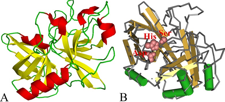
Fig 1. The predicted three-dimensional model of T. spiralis TsSP protein.

A: The predicted three-dimensional structure of TsSP protein, there are 4 α-helixes (in red), 14 β-strand (in yellow), and 19 irregular coils (in green); B: Catalytic residues Ser-His-Asp form a pocket-shaped functional domain. The active site of TsSP was highlighted with red color.
