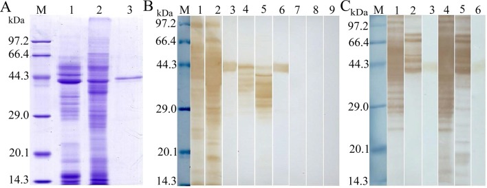
Fig 6. Western blot analysis of rTsSP antigenicity.
(A) SDS-PAGE analysis of ML ES antigens from T. spiralis (lane 1), ML crude antigen (lane 2), and rTsSP (lane 3). (B) Western blotting of the rTsSP. T. spiralis ML ES antigens (lane 1) and ML crude antigens (lane 2) and rTsSP (lane 3) were probed with mouse infection sera. The natural TsSP protein in ML ES (lane 4), ML crude antigens (lane 5) and rTsSP (lane 6) were identified by using anti-rTsSP serum. Normal mouse sera did not probe the ES (lane7) and crude antigens (lane 8), and rTsSP (lane 9). (C) Western blotting of ML ES (lane 1 and 4) and crude antigens (lane 2 and 5), and rTsSP (lane 3 and 6) probed with immune sera from mice vaccinated with ES and crude antigens.

