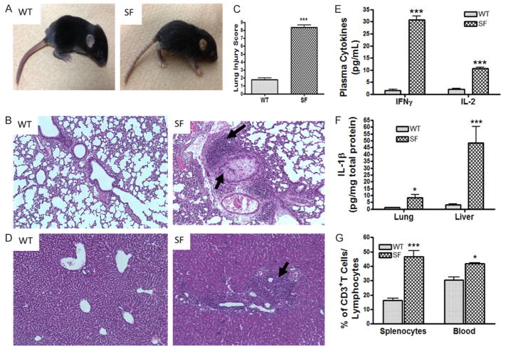
Figure 1.
Phenotypic features of scurfy mice (SF). A. A SF mouse at day 13 of life is compared to an age- matched WT mouse. It has developed scaly skin on their ears, eyes and tails, and deformed ears. B. Histology (H&E staining) of lung (100 × magnifications). Arrows indicate lymphocyte infiltrate and proteinaceous debris that filled airspaces in SF. C. Lung Injury Score (LIS) of SF compared to WT mice. D. Histology of Liver (100 × magnifications). Arrows indicate lymphocyte infiltration. E. Plasma cytokine levels of IFNγ and IL-2 in SF compared to WT. F. Inflammatory cytokine IL-1β level in lung and liver tissue lysates of SF mice compared to WT mice. G. The percentage of CD3+ T cells in the lymphocyte population from spleen and peripheral blood, comparing SF to WT mice, as analyzed by flow cytometry. All numbers represent means ± SE, *P<0.05, ***P<0.001.

