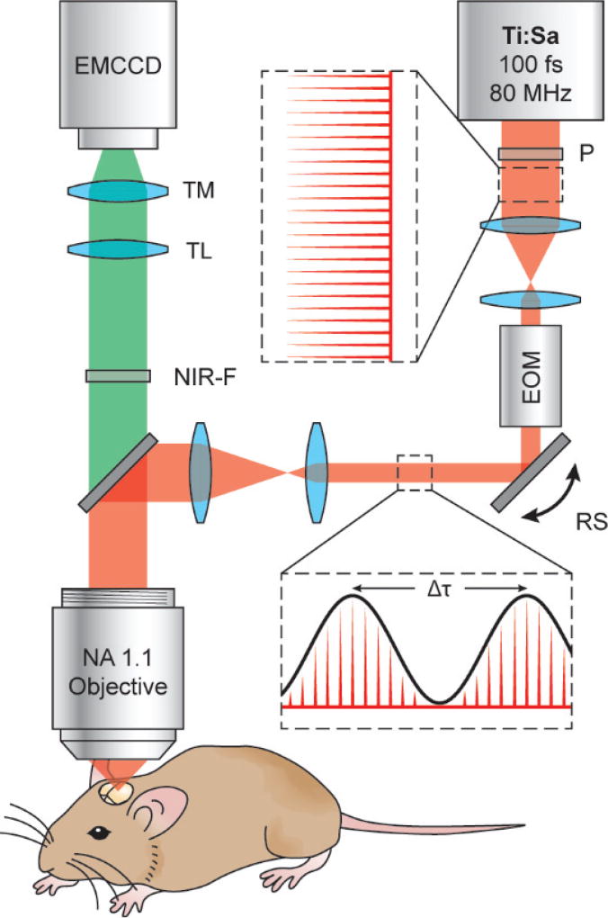
Fig. 1.
Schematic of the resonant 2P-SuPER microscopy experimental setup. The output of the laser is modulated with a frequency of 1/Δτ. The frequency is synchronized with the X-Y scan to generate a grid shaped illumination pattern at the imaging plane. Ti:Sa: titanium sapphire ultrafast laser; P: polarizer; EOM: electro-optical modulator; RS: closed loop X-Y resonant scanner assembly; NIR-F: near infrared filter; TL: tube lens; TM: tunable magnification optics; EMCCD: electron-multiplied CCD.

