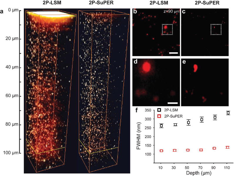
Fig. 2.
(a) 2P-LSM (left) and resonant 2P-SuPER (right) three-dimensional image of 100 nm green fluorescent nanospheres embedded in a scattering agarose gel. A comparison of images acquired by (b) 2P-LSM and (c) resonant 2P-SuPER at 90 µm from the surface of the agarose sample demonstrate the improved resolution of resonant 2P-SuPER (scale bar: 5 µm). A magnified comparison of (d) resonant 2P-LSM and (e) resonant 2P-SuPER resolution improvement (scale bar: 1 µm). (f) FWHM measurements from 10 spheres at each depth show relative independence of resolution from depth in resonant 2P-SuPER but not in 2P-LSM (error bars: sem).

