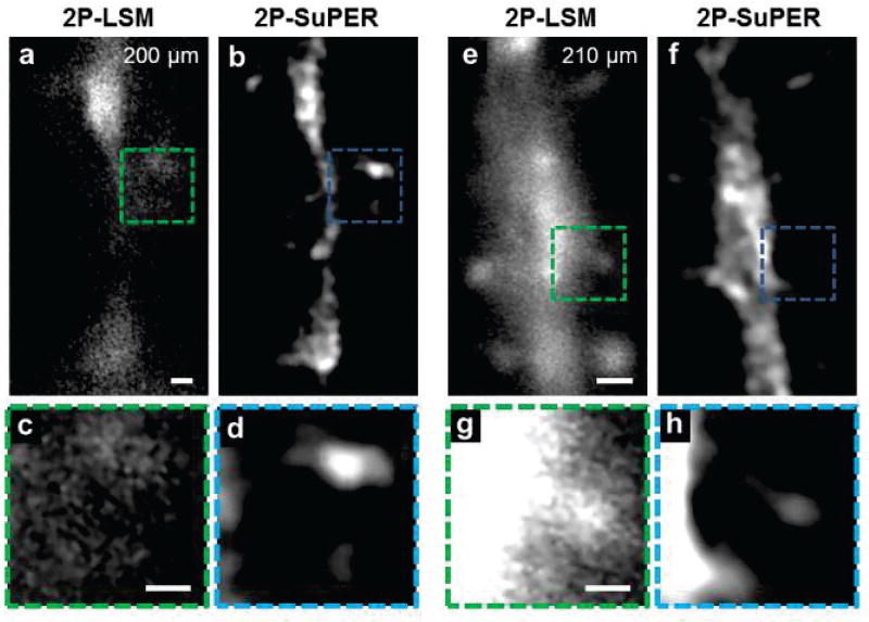
Fig. 3.
Ex vivo comparison between (a) 2P-LSM and (b) resonant 2P-SuPER imaging a eGFP-positive neuron at 200 µm in an acute brain slice at 3.5 Hz. The corresponding magnified section of the (c) 2P-LSM (green box in panels a and d) resonant 2P-SuPER (blue box in b) images show that resonant 2P-SuPER maintains super-resolution in deep tissue and is more resilient to scattering than array based 2P-LSM. Comparisons between (e) 2P-LSM and (f) resonant 2P-SuPER imaging a eGFP neurons at 210 µm in the acute brain slice at 3.5 Hz showed an analogous resolution improvement. A magnified section of the (g) 2P-LSM (green box in panels e) and (h) resonant 2P-SuPER (blue box in panel f) images show that resonant 2P-SuPER maintained super-resolution at depths up to 210 µm from the cortical surface. Scale bars: 1 µm in (a) and (e), 500 nm in (c) and (g).

