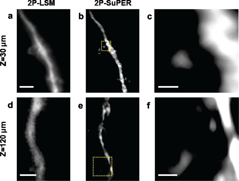
Fig. 4.
In vivo comparison between (a) 2P-LSM and (b) resonant 2P-SuPER microscopy image of a dendrite located 30 µm from the neocortical of the mouse brain and (b). (c) The magnified resonant 2P-SuPER image (indicated by the yellow box in panel b) shows the dendritic spine extending from the neuronal dendrite. Comparison between (d) 2P-LSM and resonant 2P-SuPER image of a dendrite located 120 µm from the neocortical surface. (f) The magnified resonant 2P-SuPER image (indicated by the yellow box in panel e) shows dendritic spines emerging from the neurite. Scale bars: 4 µm in (a), 1 µm in (c), 2 µm in (d), and 500 nm in (f).

