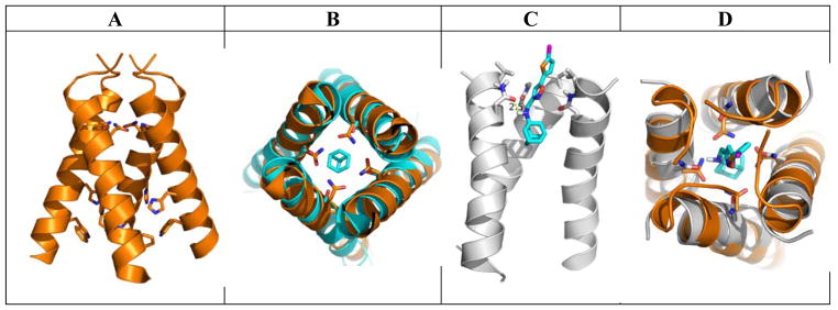
Figure 4.
Molecular docking of compound 9q in the AM2-S31N channel. (A) X-ray crystal structure of drug-free AM2-S31N structure (PDB: 3C02).22 (B) Overlay of the drug-free AM2-S31N structure (orange) and the amantadine-bound AM2-WT structure (cyan) (PDB: 3C9J).18 (C) Docking model of compound 9q in the AM2-S31N channel. The solution NMR structure (PDB: 2LY0) of AM2-S31N was used for the docking.21 Docking was performed using Autodock Vina.35 (D) Overlay of the docking model (gray) and drug-free AM2-S31N structure (orange) (PDB: 3C02).22

