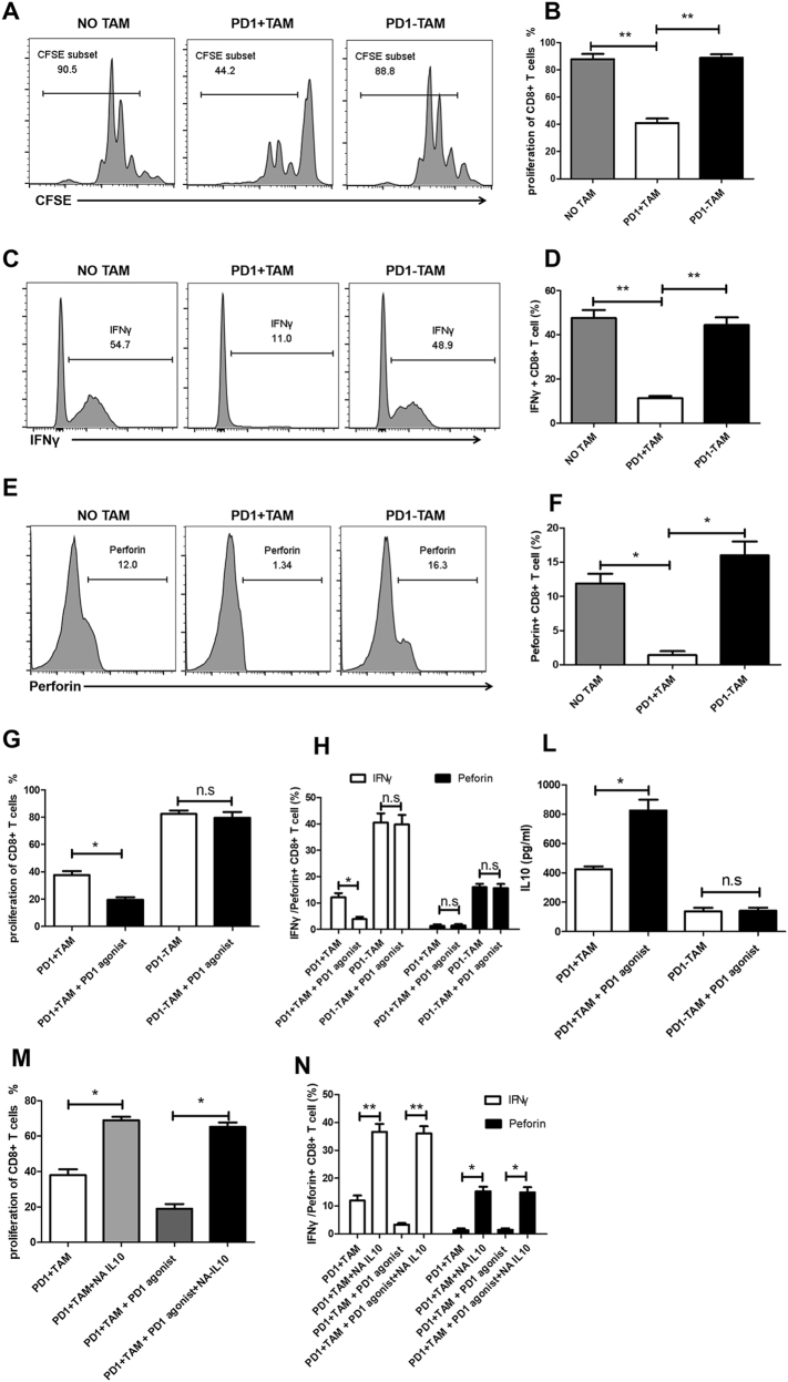
Fig. 3. PD1+ macrophages exhibit strong immunosuppressive activity.
a, b CFSE-labeled CD8+ T cells were activated by anti-CD3/CD28 beads, and PD1−, and PD1+ macrophages derived from GC tissue were added at 1:1 ratio. After 3 days, these cells were stained with anti-CD8 Ab, and the proliferation of CD8+ T cells in NO TAM (CD8+ T cells were cultured alone), PD1+ TAM (CD8+ T cells were cocultured with PD1+ macrophages), and PD1− TAM (CD8+ T cells were cocultured with PD1– macrophages) was analyzed. Representative flow cytometry plots are shown in (a). b Results represent the mean of three independent experiments. c, d Isolated CD8+T cells were activated by anti-CD3/CD28 beads, and PD1–, and PD1+ macrophages derived from GC tissue were added at 1:1 ratio. After 3 days, these cells were labeled with anti-CD8 and IFN-γAbs, and the expression of IFN-γ in CD8+ T cells in NO TAM, PD1+ TAM, and PD1– TAM was analyzed. Representative flow cytometry plots are shown in c. d Results represent the mean of three independent experiments. e, f Isolated CD8+T cells were activated by anti-CD3/CD28 beads, and PD1–, and PD1+ macrophages derived from GC tissue were added at 1:1 ratio. After 3 days, these cells were collected and stained with anti-CD8 and perforin antibody, and the expression of perforin in CD8+ T cells in NO TAM, PD1+ TAM, and PD1– TAM was analyzed. Representative flow cytometry plots are shown in (e). f Results represent the mean of three independent experiments. g, l Analysis of suppressive activity by PD1+ macrophages left untreated or incubated with 10 μg/ml anti-PD1 antibody, or polyclonal goat IgG. g CD8+T-cell proliferation was analyzed, and the data indicate the mean ± standard error of the mean of three independent experiments. h The expression of Perforin and IFNγ in CD8+T cells was analyzed, and the data indicate the mean ± standard error of the mean of three independent experiments. l the expression of IL10 in PD1+ macrophages was analyzed, and the data indicate the mean ± standard error of the mean of three independent experiments. m,n analysis of suppressive activity by PD1+ macrophages treated with NA-IL10 in the presence of 10 μg/ml anti-PD1antibody, or polyclonal goat IgG. m CD8+T-cell proliferation was analyzed, and the data indicate the mean ± standard error of the mean of three independent experiments. n the expression of Perforin and IFNγ inCD8+T cells was analyzed, and the data indicate the mean ± standard error of the mean of three independent experiments

