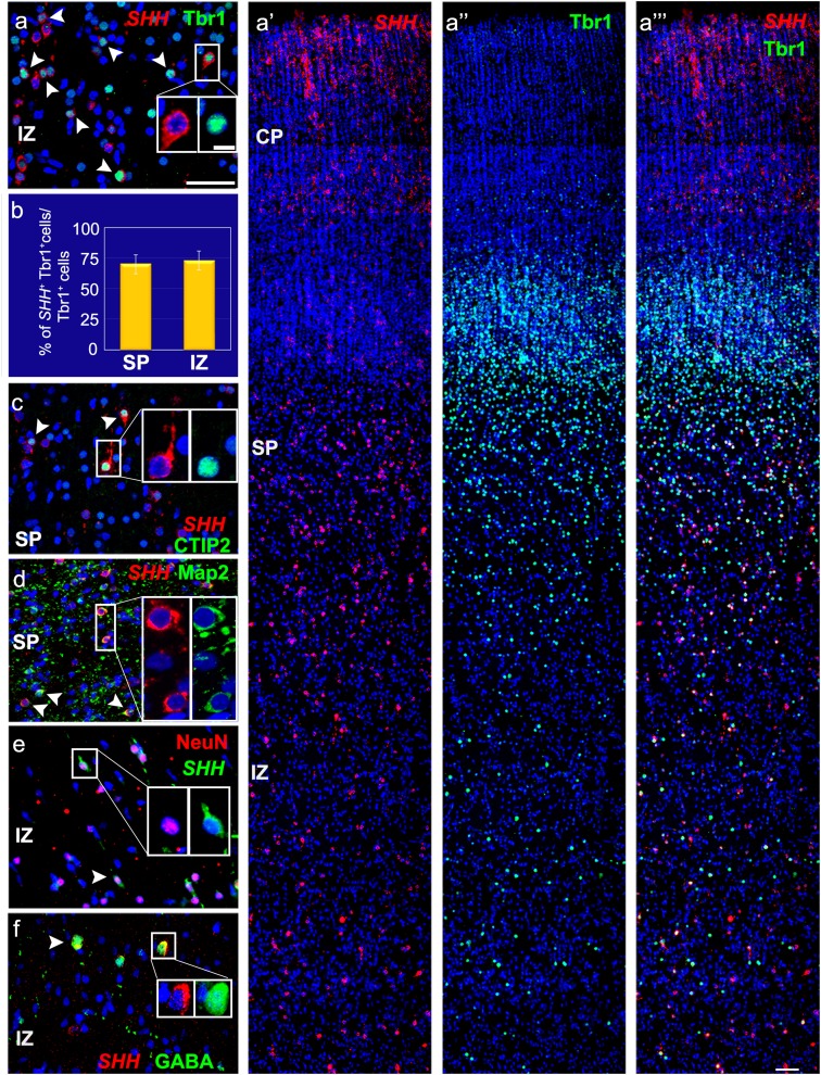
Fig. 5.
Various subtypes of neurons express SHH in the 18–24 gw cortex. a–a‴ Co-labeling for SHH mRNA (red) and Tbr1 (green) reveals many postmitotic projection neurons across the SP and IZ expressing SHH. a′, a″ Single channels. b Percentage of Tbr1+ cells expressing SHH in the SP (70% ±7.6 SEM) and IZ (73% ±7.8 SEM) in 22–24-gw sections (n = 3). c–f SHH is expressed by neurons labeled with CTIP2 (c), MAP2 (d), NeuN (e), and GABA (f). Scale bars: a and a‴ 50 µm, a-inset 10 µm

