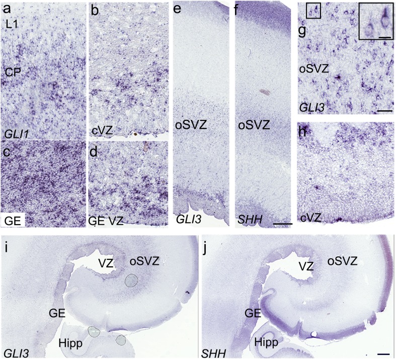
Fig. 6.
Components of the SHH-signaling pathway are expressed in the human fetal cortex at 22 gw. a, b ISH reveals the cortical expression of GLI1 (CP and VZ), indicative of SHH-signaling activity in these areas. c, d Strong GLI1 expression in the GE suggests higher levels of SHH activity ventrally. e, f GLI3 and SHH transcripts show similar high-density signals in the VZ and oSVZ. g On higher magnification, GLI3+cells in the oSVZ resemble RGCs and are more numerous than in the VZ (h). i, j Comparison of the GLI3 and SHH expression patterns in adjacent coronal sections at the level of the hippocampus (Hipp) of the 22 gw fetal brain. Scale bars: f 500 µm, g 50 µm, g- inset 15 µm, j 1 mm

