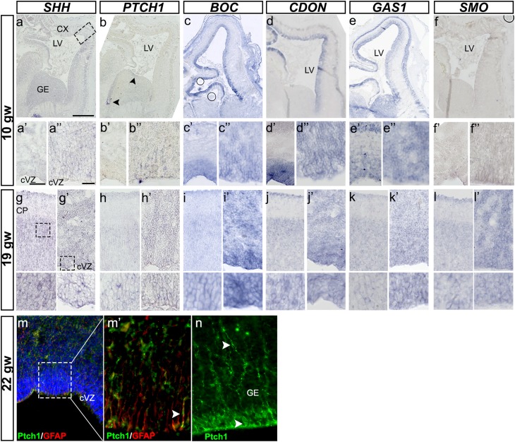
Fig. 7.
Expression of SHH receptors in the fetal human brain at 10 and 19 gw. a–a″ ISH for SHH on a coronal section from a 10-gw brain, as a reference for all SHH receptors in sections from the same fetus. Higher magnification of the boxed areas in (a): a′ cortex, a″ cVZ. Weak signal is detected in the cortical VZ and GE. b–b″ ISH for PTCH1 receptor reveals very low expression in the VZ (b″). BOC expression is much more prominent along the cortical VZ and GE VZ (c–c″), whereas CDON expression is restricted to the cortical VZ (d–d″). GAS1 is the only receptor expressed in both the VZ and CP, in addition to the GE (e–e″). SMOOTHENED (SMO) is hardly detected in the VZ at this stage (f–f″). g–l′ SHH and all its receptors in contiguous sections of 19-gw brain. Two areas are presented for each gene: CP and VZ/SVZ. m Co-staining of 22-gw brain with Patched1 and GFAP antibodies shows that RGCs in the cortical VZ express this receptor. m′ Higher magnification of the boxed area in (m). n Ptch1 immunoreaction in the same section reveals a strong expression in the GE. a 1 mm, a′ 150 µm, a″ 25 µm

