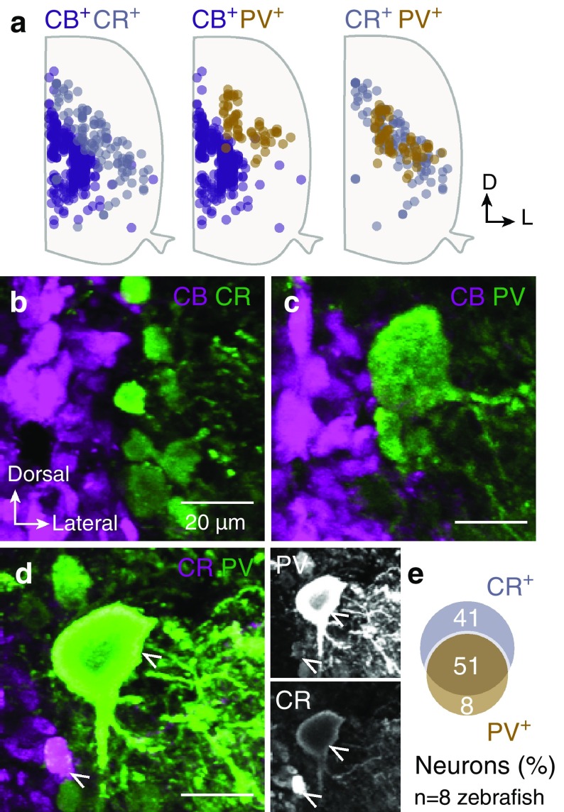
Fig. 2.
Co-distribution and co-localization of the CBPs positive neurons. a Superimposed positions of the CR, CB and PV positive neurons in the spinal cord. b–d Double immunofluorescent images between the three studied CBPs (CR, CB and PV). Many double expressing neurons observed only between CR and PV (51%; arrowheads in d). e Percentage of co-expression of CR, CB or PV in neurons. Only a small population of PV+ neurons does not express CR (8%; e). Single channel views of the respective framed box

