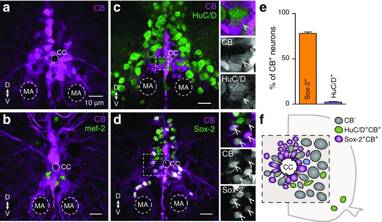
Fig. 6.
Identity of the CB positive neurons. a Distribution pattern of CB+ expression around the central canal in the adult zebrafish spinal cord. b None of the CB expressing cells (magenta) co-localized with the early neuronal differentiated marker mef-2 (green). c–d A small number (2.32%) of the differentiated and mature neurons (HuC/D+, green) contain also CB (magenta). In addition, the vast majority (78.13%) of CB containing cells (magenta) is progenitor cells / stem cells (Sox-2+, green). Single channel views shown for areas are indicated by framed rectangles. Arrowheads indicate double labeled cells. e Quantification of CB positive cells that co-express Sox-2+ or HuC/D+. f Schematic representation of the central canal area in the adult zebrafish spinal cord showing that most of the CB+ cells are progenitors / stem cells

