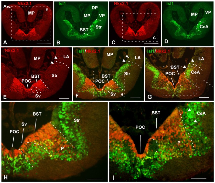
Figure 3.
Isl1/Nkx2.1 expression in the subpallium of Pleurodeles waltl. Photomicrographs of transverse sections through the telencephalon of Pleurodeles waltl at caudal levels of the interhemispheric connection of the lateral ventricles. In the subpallial part of the telencephalon, the combined immunohistochemical detection of Isl1 (green) and Nkx2.1 (red), allowed the identification of the boundaries of this region and the identification of the marked areas. Note the distribution of Nkx2.1 cells in ventricular and mantle zones, in the ventromedial region, whereas the Isl1 labeling is restricted to cells in the mantle of the lateral and medial parts of the subpallium. Arrowheads point to Nkx2.1 labeled cells in striatal, central amygdala (CeA) and ventral pallial areas. Scale bars = 500 μm (A–D), 200 μm (E–G) and 100 μm (H,I). See list for abbreviations.

