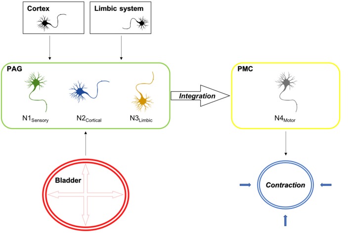
Figure 9.
Electrical stimulation mimicking stretch of the detrusor muscle leads to the activation of sensory bladder afferents (Aδ fibers), which in turn activate sensory neurons in the PAG (N1 sensory). Besides, the PAG receives cortical and limbic input, which evaluates external circumstances, which either inhibit or facilitate voiding and activate cortical (N2 cortical) and limbic (N3 limbic) projection neurons in the PAG. The integration of these signals takes places in neurons projecting to the PMC. In turn the PAG releases its disinhibitory action on the PMC, which results in the activation of efferent neurons (N4 motor), which result in the corresponding action at the spinal cord, leading to detrusor contractions and the relaxation of the external urethral sphincter, which are prerequisites for voiding. Also shown is the activation pattern as assessed by cFos expression in the D-N configuration for the rostral and caudal PAG. Red areas indicate activation and green areas are not activated upon sensory afferent stimulation.

