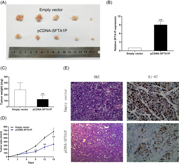
Figure 5. SFTA1P overexpression inhibits tumorigenesis of GC cells in vivo.
(A) Empty vector or pCDNA-SFTA1P were transfected into SGC7901 cells, which were injected in the nude mice (n=5), respectively. Tumors formed in pCDNA-SFTA1P group were dramatically smaller than the control group. (B) qRT-PCR was performed to detect the average expression of SFTA1P in xenograft tumors. (C) Tumor weights were represented as means of tumor weights ± S.D. (D) Tumor volumes were calculated after injection every 3 days. Points, mean (n=5); bars indicate S.D. (E) The tumor sections were under H&E staining and immunohistochemical (IHC) staining using antibodies against Ki-67. Error bars indicate mean ± S.E.M. *P-value <0.05, **P-value <0.01.

