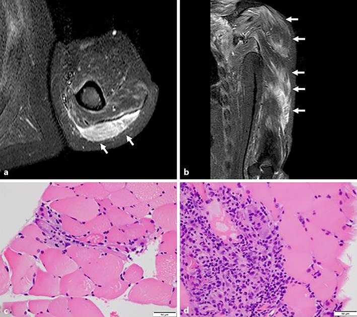
Fig. 1.

a, b Brachium magnetic resonance imaging showed increased signal intensity in the left triceps and deltoid muscles on short tau inversion recovery imaging (arrows). c, d Muscle biopsy examination of the left biceps brachii muscle revealed perifascicular atrophication and inflammatory myopathy (hematoxylin and eosin staining).
