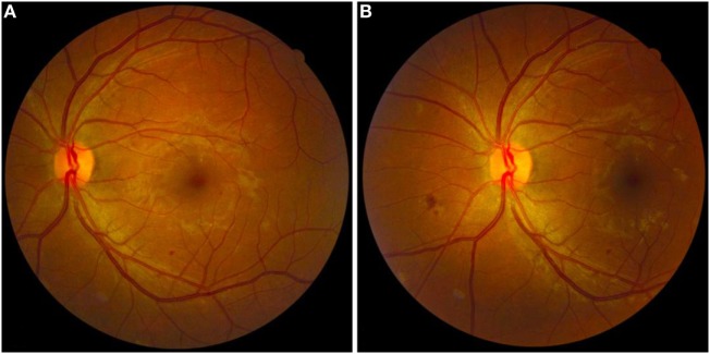
Figure 1.
Fundus retinal images of the left eye of a patient with diabetic retinopathy. (A) Fundus retinography centered on the fovea. (B) Fundus retinography centered on the optic disk. Note the presence of small hard exudate, microaneurysms, and dot-blot retinal hemorrhages around macular area (A) and small cotton wool spots and superficial retinal hemorrhages in the nasal area of the optic disk (B). The pictured eye was graded as “non-proliferative” by the retinal specialist.

