Figure 2.
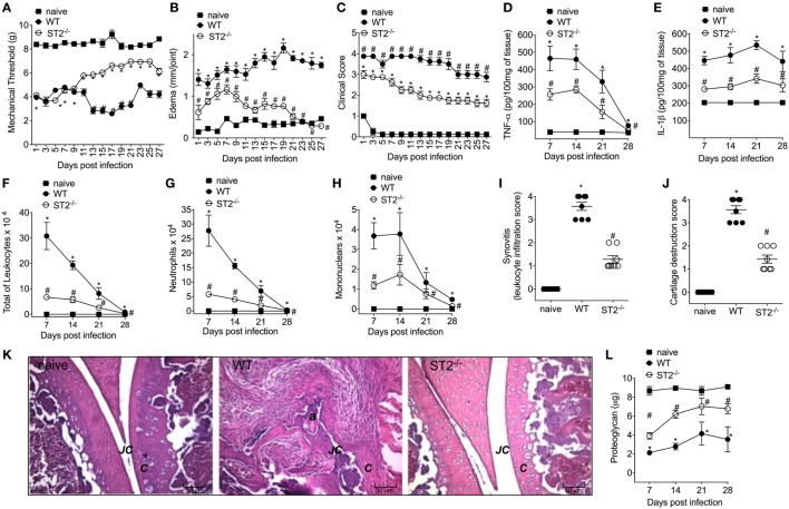
ST2 deficiency ameliorates Staphylococcus aureus-induced septic arthritis. S. aureus or saline was injected in the femur-tibial joint of wild-type (WT) and ST2−/− mice. (A) Mechanical hyperalgesia, (B) articular edema, and (C) clinical score were evaluated over 27 days post-infection. Knee joints were collected and processed to determine the levels of (D) TNF-α and (E) IL-1β by ELISA determined at days 7–28 days post-infection. (F) Total leukocytes, (G) neutrophil, and (H) mononuclear recruitment to the knee joint were determined at 7–28 days post-infection. Knee joint samples were collected at the 28th day post-infection for histological analysis by hematoxylin/eosin stained slices to determine: (I) synovitis score (intensity: 1–4) and (J) cartilage destruction score (intensity: 1–4). (K) Representative images of knee joints at 28 post-infection in original magnification ×10. The letter a indicates a heavily inflamed joint with cartilage destruction and pannus formation. (F) Proteoglycan content in patella determined at 7–28 days post-infection. For inflammatory parameters and proteoglycan content: n = 6 per group per in vivo experiment, representative of two independent experiments. *P < 0.05 vs naïve mice group, #P < 0.05 vs WT mice group (A–H,L). One-way ANOVA followed by Tukey’s test. For histological analysis: n = 8 per group per experiment, representative of two independent experiments. *P < 0.05 vs naïve mice group, #P < 0.05 vs WT mice group (I–K). Kruskal–Wallis test followed by Dunn’s test. Abbreviations: C, cartilage; JC, joint cavity.

