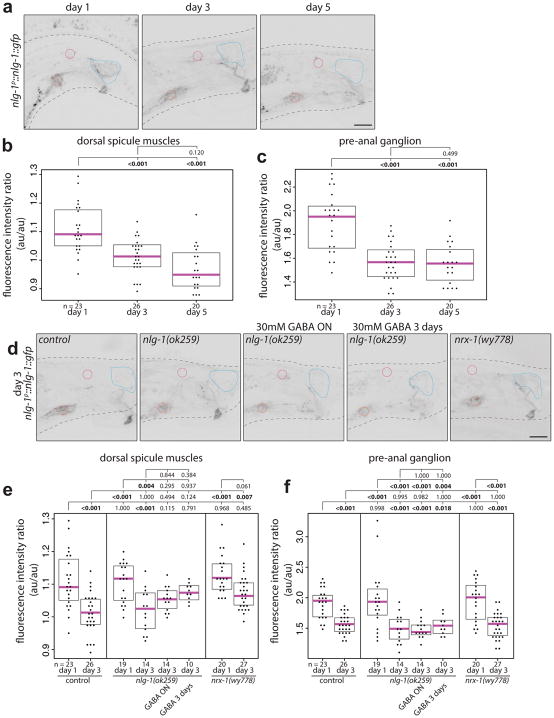
Extended Data Fig. 8. NLG-1 expression decreases from day 1 to day 3.
(a) Confocal images of nlg-1p::nlg-1::gfp in males at day 1, 3, and 5. Example regions of interest for measurements taken from single planes – blue – dorsal spicule muscles, red – pre-anal ganglion, magenta – DVB. Quantification of fluorescence intensity of nlg-1p::nlg-1::gfp in males at day 1, 3, and 5 reported as a ratio of mean fluorescence in (b) dorsal spicule muscles or (c) pre-anal ganglion normalized to background of DVB, which has little to undetectable expression. Dorsal spicule muscles refer to the gubernacular retractor, gubernacular erector, anterior oblique, anal depressor. (d) Confocal images of nlg-1p::nlg-1::gfp in control, nlg-1(ok259), nlg-1(ok259) with overnight GABA exposure, nlg-1(ok259) with 3 day GABA exposure, and nrx-1(wy778) males at day 3. Quantification of fluorescence intensity of nlg-1p::nlg-1::gfp in day 1 and 3 control, nlg-1(ok259), and nrx-1(wy778) males and day 3 nlg-1(ok259) with overnight GABA exposure and nlg-1(ok259) with 3 day GABA exposure, as a ratio of mean fluorescence in (e) dorsal spicule muscles or (f) pre-anal ganglion normalized to background of DVB. (dot=one animal, magenta bar=median, and boxes=quartiles, one-way ANOVA and post-hoc Tukey HSD).

