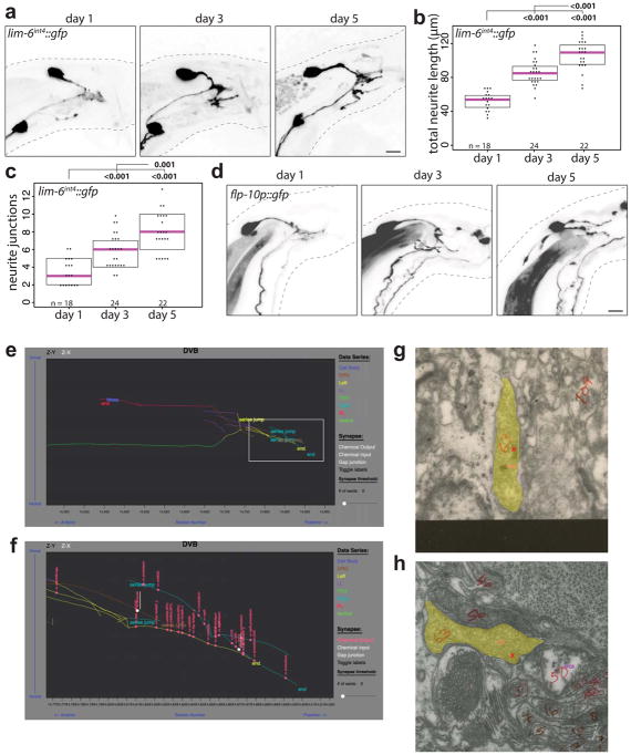
Extended Data Fig. 1. Progressive neurite outgrowth in DVB in adulthood.
(a) DVB neuron visualized with lim-6int4::gfp at days 1, 3, and 5 in adult males and quantified by (b) total neurite length and (c) number of neurite junctions (dot=one animal, magenta bar=median, and boxes=quartiles, one-way ANOVA and post-hoc Tukey HSD). (d) DVB neurite outgrowth visualized with flp-10::gfp in males at days 1, 3, and 5 of adulthood (n>10). (e) Tracing reconstruction of male DVB from EM sections compiled by wormwiring.org showing DVB neurites. (f) Inset of DVB neurites showing pre-synaptic specializations identified in EM sections shown in pink. EM section showing DVB pseudo-colored yellow with pre-synaptic specialization indicated with red ‘x’ with (g) SPCR (Image Right1200, Section 14871) and (h) spicule sheath (Image N2YDRG1175, Section 14816), shown in white in inset panel.

