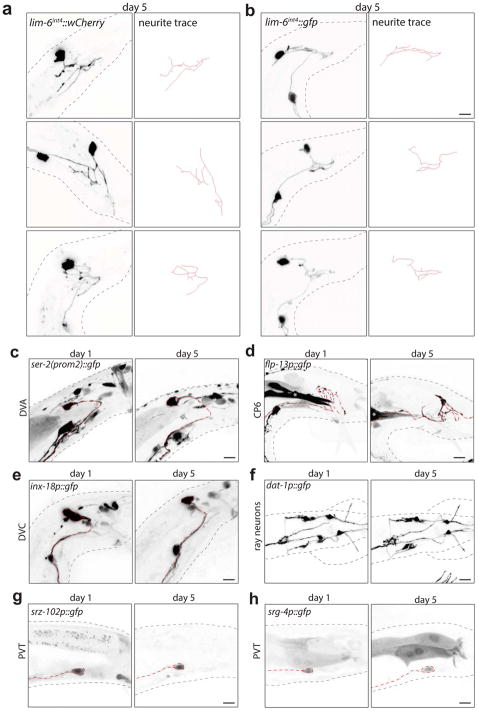
Extended Data Fig. 2. DVB neurite outgrowth in adult male C. elegans is stochastic and other neurons in the male tail do not show progressive neurite outgrowth in adulthood.
DVB neurites at day 5 visualized with (a) lim-6int4::wCherry or (b) lim-6int4::gfp (n>10 for each). DVB posterior neurites were traced through confocal stacks using Simple Neurite Tracer4 plugin. (c) DVA neuron visualized with ser-2(prom-2)::gfp (n=5)(red dashed line indicates axon of relevant neuron), (d) DVC neuron visualized with inx-18p::gfp (n=5), (e) CP6 neuron visualized with flp-13::gfp (cell soma not shown) (n=5), (f) ray neurons visualized with dat-1::gfp (ventral view)(n=5). PVT neuron visualized with srz-102p::gfp (n=5)(g) and srg-4p::gfp (n=5)(h) at day 1 and day 5. Axons of indicated neurons highlighted by red dashed line.

