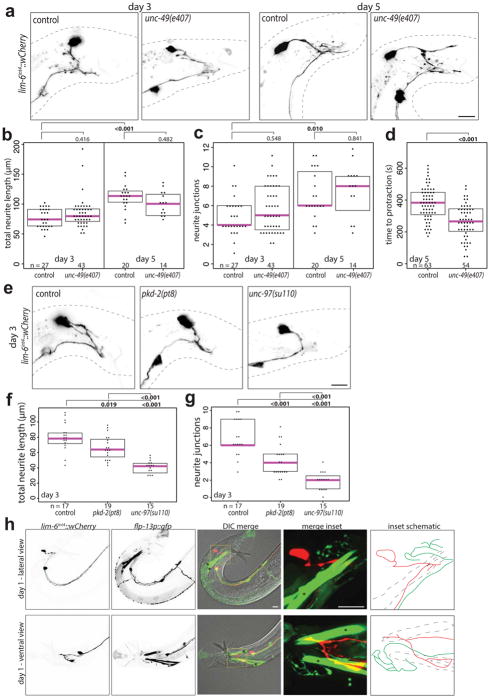
Extended Data Fig. 4. DVB neurite outgrowth in unc-49, pkd-2 and unc-97 mutant males. flp-13p::gfp labels CP6 and spicule retractor muscles.
(a) Confocal images and quantification of (b) total neurite outgrowth and (c) number of neurite junctions in control and unc-49(e407) males at days 3 and 5. (d) Time to spicule protraction on aldicarb at day 5 for control and unc-49(e407) males. (e) Confocal images and quantification of (f) total neurite outgrowth and (g) number of neurite junctions in control, pkd-2(pt8), and unc-97(su110) males at day 3. (h) Confocal images of male worms with lim-6int4::wCherry, flp-10p::gfp, and DIC at day 1 in ventral and lateral views. Inset showing DVB and CP6 axons, with schematic of axons demonstrating lack of contact (red is DVB axon, green is CP6 axon, blue dashed lines are spicule retractor muscles). Asterisks in flp-13::gfp panel mark spicule retractor muscles. (dot=one animal, magenta bar=median, and boxes=quartiles, one-way ANOVA and post-hoc Tukey HSD).

