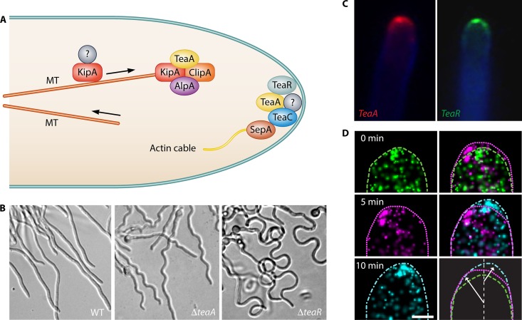
FIG 3.
Cell end marker proteins determine the interplay between the microtubule and the actin cytoskeleton in A. nidulans. (A) Scheme of cell end markers transported at the MT plus end and delivered to the apical membrane. The prenylated TeaR proteins are probably delivered with vesicles. The motor protein KipA transports TeaA and probably other tip proteins toward the MT plus end. (B) Differential interference contrast images of wild-type, ΔteaA, and ΔteaR strains. ΔteaA strains exhibited zigzag and ΔteaR strains curved hyphae. (C) Monomeric red fluorescent protein 1 (mRFP1)-TeaA or GFP-TeaR localizes to one point at the tip and along the tip membrane. (D) Series of PALM images of an mEosFP-TeaR-expressing hypha (5-min time interval). Cell profiles are shown in different line styles. The right column shows overlays of PALM images from two time points (top, 0 and 5 min; middle, 5 and 10 min), and overlays of outlines reveal growth regions coinciding with TeaR cluster locations. (Panels A to C are modified from reference 64 with permission; panel D is modified from reference 77.)

