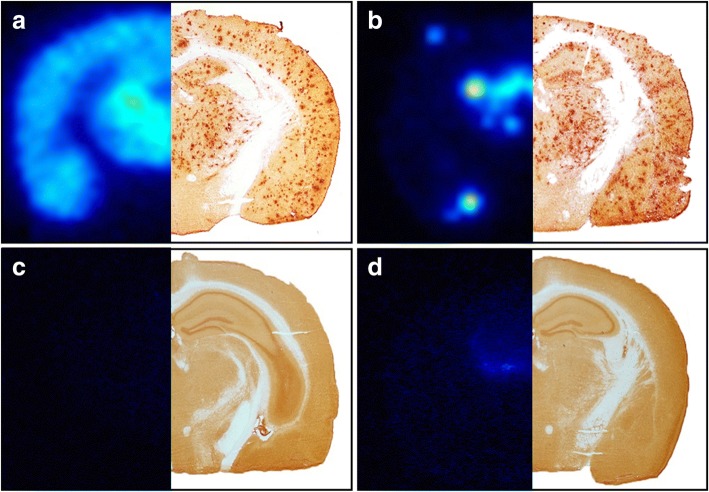
Fig. 1.
Global antibody distribution in the brain. Brain distribution in 18-month-old tg-ArcSwe mice given an injection with [125I]RmAb158-scFv8D3 (a) and [125I]RmAb158 (b), 6 days postinjection, as visualized with ex vivo autoradiography (left) in comparison with amyloid-β 40 immunostaining (right). Whereas [125I]RmAb158-scFv8D3 was distributed throughout the whole brain, [125I]RmAb158 was confined to central parts of the brain. For comparison, also wild-type animals were given an injection with [125I]RmAb158-scFv8D3, resulting in no signal (c) and with [125I]RmAb158, where a faint signal was detected centrally in the brain (d)

