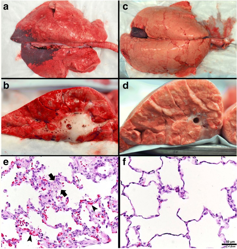
Fig. 8.

Gross Lung Photos: The top panel a-d shows gross photos of the whole lung and the lung cut surface at necropsy in a clinically applicable 48-h peritoneal sepsis plus gut ischemia/reperfusion, porcine, acute respiratory distress syndrome (ARDS) model. The lungs were inflated to 25 cmH2O when photographed, to standardized lung volume history (top panel a, c). One group of animals was place on the ARDSnet protocol immediately following injury (top panel a, b). The other group was placed on the time-controlled adaptive ventilation (TCAV) protocol immediately following injury (top panel c, d). Preemptive application of the ARDSnet protocol did not prevent the development of ARDS. A large area of consolidation (dark red), inflammation (reddish color), and a lung not fully inflated at an airway pressure of 25 cmH2O is shown (top panel a). The cut lung surface also demonstrated inflammation throughout the lung tissue and copious edema foam flowing from the large airways (top panel b). The preemptive TCAV protocol prevented the development of ARDS with the lung appearing pink (no inflammation) and fully inflated (top panel c). Inflated pink tissue was seen throughout the cut lung surface and no edema foam was seen in the airways (top panel d). Lung Histology:The bottom panel shows representative histology staining in the ARDSnet (e) and TCAV protocol (f). Lung tissue from the ARDSnet protocol group showed alveolar wall thickness (between arrows) and vessel congestion (arrowheads) (bottom panel e), which were not seen in the TCAV protocol group (bottom panel f) [5]
