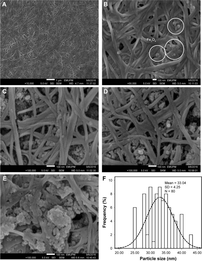
Figure 5.
FESEM images of pure (A) BNC and (B–E) BNC/Fe3O4 nanocomposites (1.0, 4.0, 8.0, and 16.0 wt%), respectively, and (F) particle size distribution of Fe3O4 NPs.
Abbreviations: FESEM, field emission scanning electron microscopy; BNC, bacterial nanocellulose; NPs, nanoparticles; SEI, upper detector; WD, working distance between the sample surface and the low portion of the lens; EMUPM, electron microscope of Universiti Putra Malaysia.

