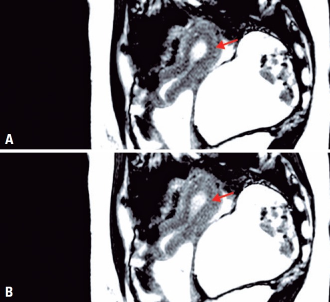
Figure 1. Uterine pelvis, cine-RM sequence on sagittal plane, with focal contraction uterine fundus. (A) Uterus without contraction, note homogeneous, regular junctional zone in the posterior aspect of the fundus. (B) Uterus with contraction, note imprint/narrowing of the junctional zone in the posterior aspect of the fundus, as it becomes thinner in response to uterine peristalsis.

