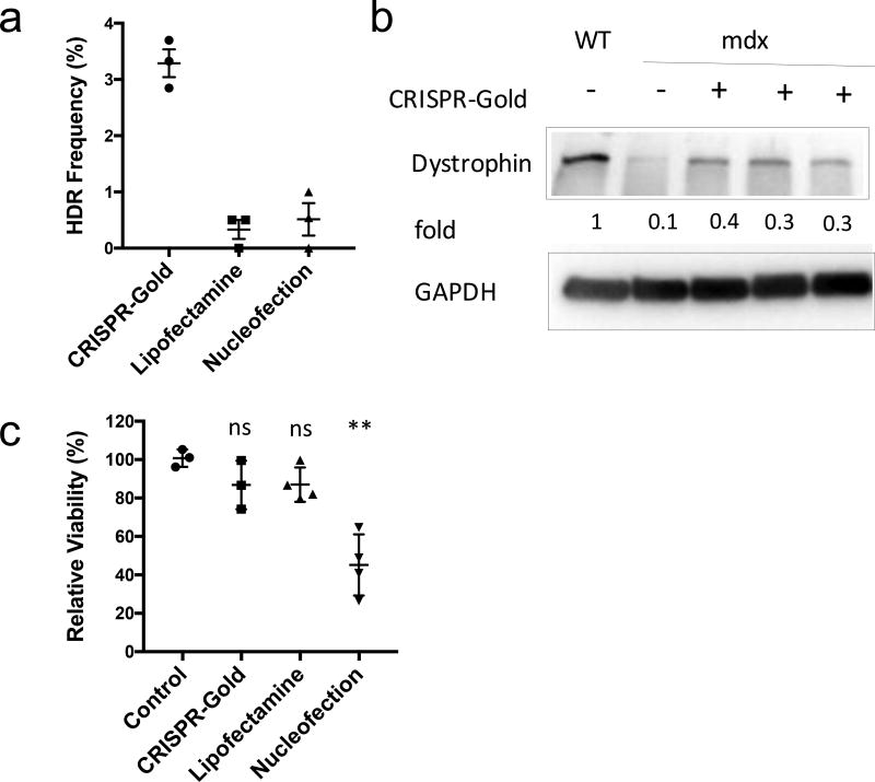
Figure 4. CRISPR-Gold induces HDR and promotes the expression of dystrophin protein in primary myoblasts.
a) The dystrophin gene was edited with CRISPR-Gold in primary myoblasts from the mdx mouse. CRISPR-Gold corrected the nonsense mutation in the dystrophin gene of mdx myoblasts with an HDR efficiency of 3.3%, which is significantly higher than either nucleofection or lipofectamine. No correction was observed in the negative control, composed of CRISPR-Gold without gRNA (data not shown). Mean ± S.E, n=3. A one-way ANOVA test had a p= 0.0002.
b) Dystrophin protein is expressed in myotubes that were differentiated from CRISPR-Gold edited primary mdx myoblasts. Western blot analysis was conducted to quantify the levels of dystrophin protein in muscle cells. The fold expression was determined by dividing the band pixel density of each group with the dystrophin band intensity from WT (C57.B6) myotubes (n=1). GAPDH was used as a loading control.
c) CRISPR-Gold causes minimal toxicity to primary myoblasts, whereas nucleofection caused significant toxicity. Primary mdx myoblasts were transfected with the indicated methods and cell viability was measured 2 days after the transfections with the cell counting kit-8. Relative viability to control, mean ± S.E, n=6. *, p < 0.05, ns = statistically not significant to control.

