Figure 4.
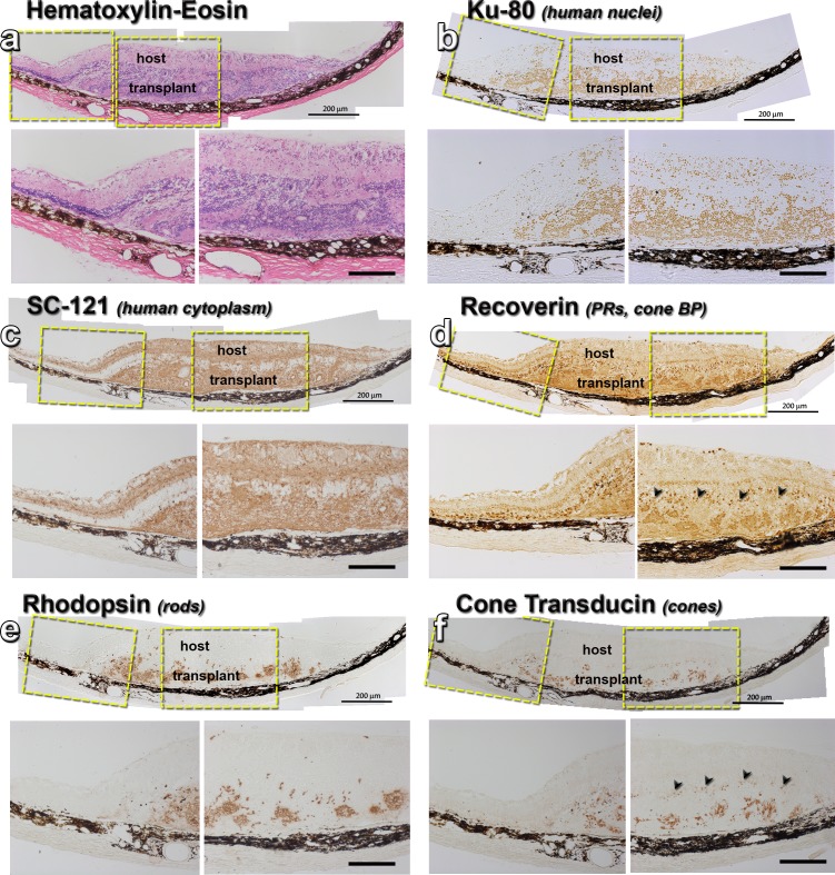
Histology of transplant no. 7 with best SC response. Each panel shows an overview of the transplant area, with enlargements of the transplant-host interface and main transplant area underneath. (a) Hematoxylin-eosin staining of a 183 dps transplant in the subretinal space of an RD rat. (b) Ku80 stain (human nuclei) stains the transplant. Some donor cells have migrated into the host retina. (c) SC121 staining (human cytoplasm) shows donor processes extending into host retina. (d) Recoverin (photoreceptors, cone bipolar cells) staining of transplant rosettes. There are some remaining host recoverin+ cones and they are highlighted by black arrows in the enlargement. (e) Rhodopsin staining of transplant rosettes. No stain in host. (f) Cone transducin staining of transplant rosettes. Only very faint, scattered stain (black arrows) in host. Scale bars: 200 μm.

