Figure 1.

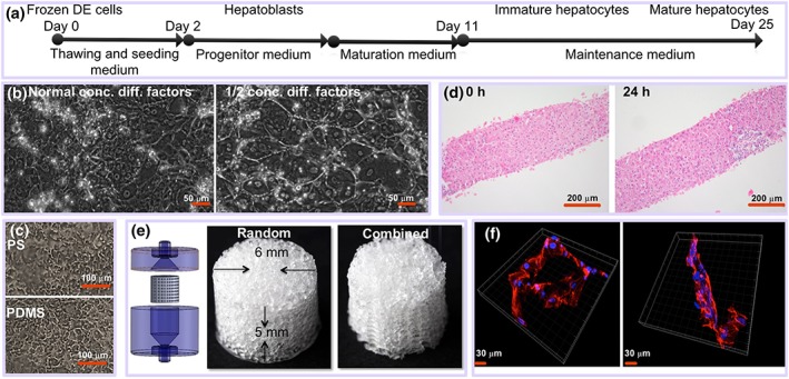
(a) Schematic overview of the experimental process of differentiation of DE cells to mature hepatocytes. (b) Differentiation of induced pluripotent stem cell (iPSC) under flow regime (500 nl/min) on flat surface for 25 days using normal concentration of differentiation factors or ½ concentration of differentiation factors. (c) Differentiation of iPSC for 19 days on polystyrene (PS) and on polydimethylsiloxane (PDMS). (d) Human precision‐cut liver slices at 0 and 24 hr of incubation (haematoxylin–eosin staining). (e) Scaffolds and house (left panel) used for perfusion. (f) Confocal microscopy images of iPSCS‐derived hepatocyte‐like cells. Blue is nucleus (DAPI), and red is actin staining [Colour figure can be viewed at http://wileyonlinelibrary.com]
