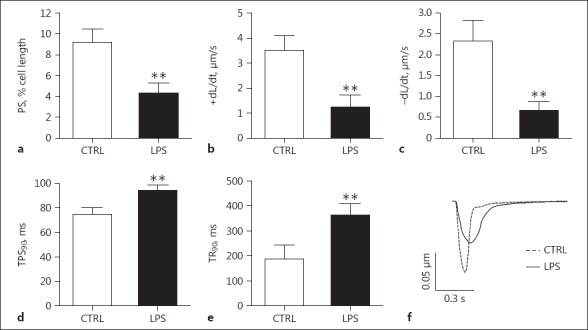
Fig. 3.

Contractile function of single cardiomyocytes. Cardiomyocytes were isolated from the hearts of the septic rats euthanized at 4 h after LPS treatment. a Peak shortening (PS). b Peak rate of shortening (+dL/dt). c Peak rate of relengthenin (–dL/dt). d Time to 90% peak shortening (TPS90). e Time to 90% of relengthening (TR90). f Representative traces of cellular sarcomere shortening in rat LV myocytes paced at 1 Hz. n = 88 from 5 LPS-treated mice; n = 35 from 5 control mice. ** p < 0.01 compared to the control.
