
Fig. 5.
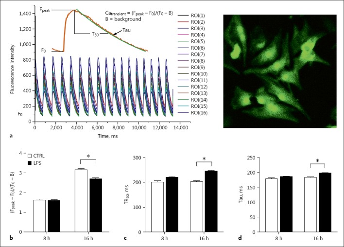
Measurements of intracellular Ca2+ transients in neonatal cardiomyocytes. The cells were exposed to LPS for 8 or 16 h. a A demonstration of Ca2+ transient of multiple cells (left) and a representative view of cultured rat neonatal cardiomyocytes under ×40 magnification (right). b Amplitude of Ca2+ transient (Fpeak – F0/F0 – B). c Time to 50% Ca2+ decay (TR50). d Time constant of Ca2+ decay (Tau). n = 130–150. * p < 0.05 compared to the control.