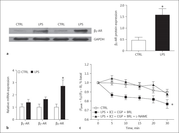
Fig. 6.

The expression of β-ARs in rat neonatal cardiomyocytes with 16 h of LPS treatment. a The expression level of β3-AR protein detected by immunoblotting. A representative immunoblotting image (left) and the quantitative data (right). n = 4. b The expression levels of β1-, β2-, and β3-AR mRNA. n = 4. c The amplitude of Ca2+ transients in the LPS-treated cardiomyocytes with addition of the following compounds. BRL 37344 (0.1 μM) is a β3-AR agonist. CGP 20712 (0.3 μM) is a specific β1-AR antagonist. ICI 118551 (0.1 μM) is a specific β2-AR antagonist. L-NAME (10 μM) is a NOS inhibitor. The Ca2+ transient was measured at 0, 5, 10, 15, 20, and 30 min after the addition of the β3-AR agonist. n = 40–45. * p < 0.05 versus the control.
