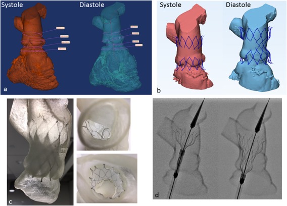
Figure 3.

Pre‐procedural evaluation. A, Three‐dimensional CT reconstruction of the right heart in systole and diastole used to calculate RVOT dimensions including lengths, diameters, and circumference along the length of the potential deployment zone. B, Virtual “implants” of an unconstrained, fully expanded device in systole and diastole examining the extent of device contact with vessel wall. C, Deployments of the device within a three‐dimensional soft rubber model of the RVOT. D, Still fluoroscopic frames of a test deployment using the same three‐dimensional model.
