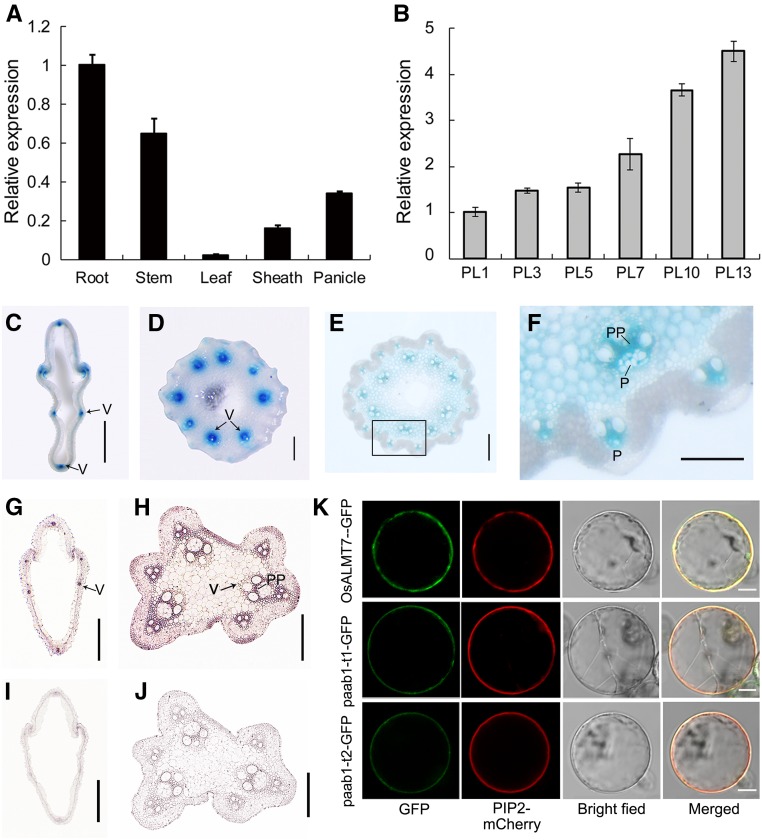
Figure 5.
Expression Pattern and Subcellular Localization of the OsALMT7 Protein.
(A) Relative expression of OsALMT7 in various tissues, including root, stem, leaf blade, leaf sheath, and panicle at the 7-cm stage (PL7). Rice UBIQUITIN was used as an internal control. Data are presented as mean ± se (n = 3).
(B) Relative expression of OsALMT7 in developing panicles at the 1-, 3-, 5-, 7-, 10-, and 13-cm stages (PL1 to PL13) before heading. Rice UBIQUITIN was used as an internal control. Data are presented as mean ± se (n = 3).
(C) to (F) Promoter activity of OsALMT7 as shown by GUS staining. Vascular tissue-preferred expression was seen in spikelet hull (C) and rachises at basal (D) and middle (E) parts of the panicles carrying the OsALMT7-GUS fusion. The squared area in (E) was magnified to show a vascular tissue (F). P, phloem; PP, phloem parenchyma cells. Bars = 0.5 mm in (C), 0.2 mm in (D) and (E), and 0.1 mm in (F).
(G) to (J) mRNA in situ hybridization of OsALMT7 on transverse sections of panicle spikelet hull (F) and rachis (G). Abundant OsALMT7 transcripts were detected in vascular tissues of the panicle. OsALMT7 sense probe was used as a negative control (I) and (J). V, vascular tissue; PP, phloem parenchyma cells. Bars = 0.5 mm in (G) and (I) and 0.2 mm in (H) and (J).
(K) Plasma membrane localization of OsALMT7 in rice protoplasts. The OsALMT7-GFP fusion protein was transiently coexpressed with PIP2-mCherry, a plasma membrane marker in rice protoplasts (upper row). cDNAs corresponding to the two mutant versions of transcripts detected in paab1-1 were also fused to GFP (paab1-t1-GFP and paab1-t2-GFP) and coexpressed with PIP2-mCherry in rice protoplasts (middle and lower rows). From left to right: image of GFP (green), mCherry (red), protoplast, and merged GFP and mCherry. Bars = 5 μm.

