
Figure 1.
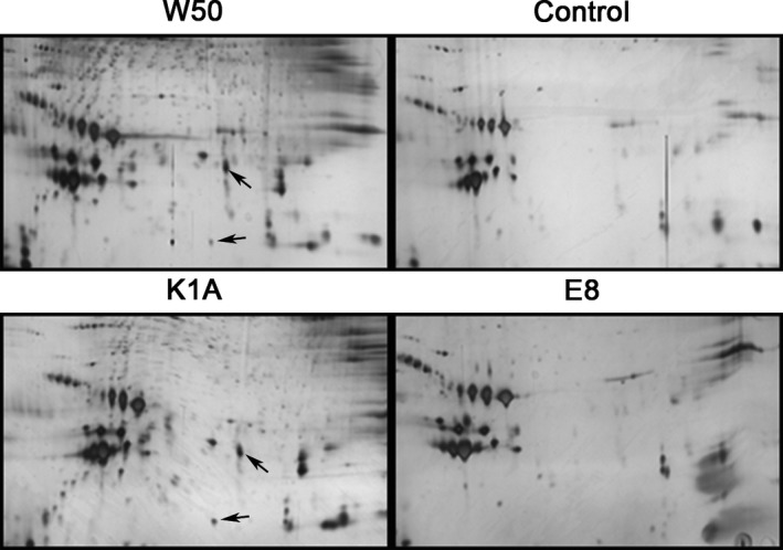
Porphyromonas gingivalis produced additional protein spots in the silver‐stained 2‐dimensional gel electrophoresis (2DE) pattern of low‐density lipoprotein/very‐low‐density lipoprotein (LDL/VLDL). The protein pattern from whole blood exposed to the W50 wild‐type strain of P. gingivalis, the gingipain mutants E8 (lacking RgpA and RgpB) and K1A (lacking Kgp) (5 × 10⁷ colony‐forming units [CFU]/mL at 37°C, 30 minutes) and the unstimulated control from the same subject are shown. Arrows indicate two non‐identified fragments