Figure 2.

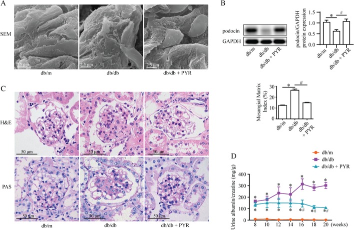
AGEs induce podocyte injury and renal tissue damage in db/db mice. (A) Representative scanning electron micrographs of the renal cortex from the indicated mice. PYR protected against abnormal podocytes in db/db mice. Scale bar = 2.5 μm. (B) Western blotting of podocin expression from the renal cortex suggesting that PYR recovered the decreased podocin level in db/db mice (mean ± SEM, n = 3). Two‐way ANOVA, followed by post hoc Student–Newman–Keuls test; *p < 0.05 versus db/m mice; # p < 0.05 versus db/db mice. (C) H & E and periodic acid–Schiff (PAS) staining of renal tissues. The mesangial matrix index indicated greater mesangial expansion in db/db mice than in non‐diabetic controls, whereas PYR reduced this mesangial expansion. Scale bar = 50 μm (means ± SEM, n = 5; 30 images from each group). Two‐way ANOVA, followed by post hoc Student–Newman–Keuls test; *p < 0.05 versus db/m mice; # p < 0.05 versus db/db mice. (D) Urinary albumin excretion (mg/g creatinine) of db/m, db/db, and db/db + PYR mice. Urine samples were collected in a metabolic cage at 8, 10, 12, 14, 16, 18, and 20 weeks of age. PYR attenuated the increased albuminuria in db/db mice at 16–20 weeks (n = 3). Repeated measures ANOVA followed by post hoc Student–Newman–Keuls test; *p < 0.05 versus db/m mice; # p < 0.05 versus db/db mice.
