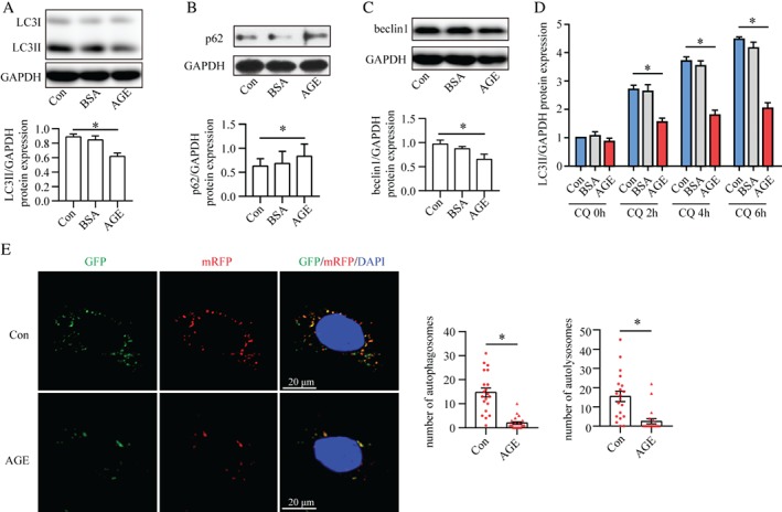
Figure 3.

AGEs impair the formation and turnover of autophagosomes. (A–C) Western blots of the expression of LC3II (A), p62 (B), and beclin1 (C) in cultured podocytes treated with vehicle (Con), BSA or AGE–BSA (AGE). Autophagic flux was inhibited in AGE‐stimulated podocytes compared with Con and BSA, as demonstrated by the reduction in expression of LC3II and belin1 as well as the accumulation of p62 (means ± SEM, n = 3–4). One‐way ANOVA, followed by post hoc Student–Newman–Keuls test; *p < 0.05 versus control. (D) AGEs reduced chloroquine (CQ)‐induced LC3II accumulation according to western blotting, suggesting blockade of autophagosome formation (mean ± SEM, n = 3). One‐way ANOVA, followed by post hoc Student–Newman–Keuls test; *p < 0.05 versus control. (E) Confocal laser scanning microscopy images of podocytes transfected with the tandem GFP‐mRFP‐LC3 adenovirus. The number of autophagosomes (yellow) and autolysosomes (red) was reduced under AGE stimulation, indicating dual suppression of the formation and turnover of autophagosomes (mean ± SEM, n = 3; 20–21 cells from each group). Student's t‐test; *p < 0.05 versus control. Scale bar = 20 μm.
