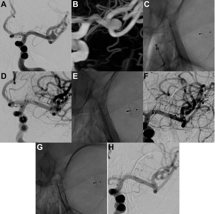
Figure 2.
Unruptured left middle cerebral artery aneurysm. (A) DSA, working view and (B) 3D-DSA show the aneurysm (transverse diameter: 6.3 mm; height: 5.3 mm; neck: 5.4 mm). (C) and (D) DSA at the end of the procedure (unsubtracted and subtracted view, respectively) show the detached WEB device (WEB SL 7×3 mm) and residual flow in the aneurysm and the device. (E) and (F) DSA at 6 months (unsubtracted and subtracted views, respectively) show the WEB device and complete aneurysm occlusion. (G) and (H) DSA at 12 months (unsubtracted and subtracted views, respectively) shows the WEB device and stable complete aneurysm occlusion.

