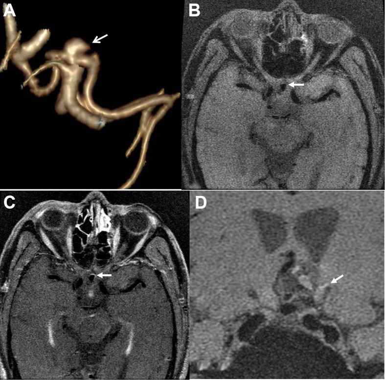
Figure 3.

A middle-aged man presented with headache due to an aneurysm in the anterior communicating artery. Three-dimensional volume rendering (VR) by CT angiography shows the irregular shape of the aneurysm (arrow) (A). Post-contrast MRI (C) indicates partial wall enhancement (arrows) compared with the pre-contrast image (B). Nineteen days later, the aneurysm ruptured with subarachnoid hemorrhage (arrow) (D).
