
Figure 4.
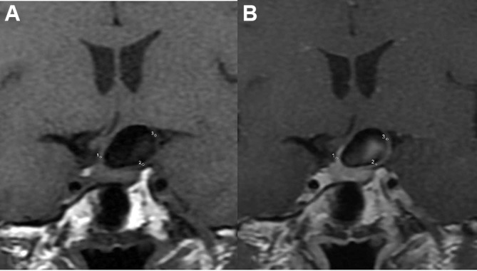
Signal intensity at the neck, body, and dome portions of the intracranial aneurysms was measured manually on the pre- (A) and post-enhancement (B) images. The signal intensity values were determined in circular regions of interest placed in the aneurysm wall, and average values were used.