. 2018 Apr 24;9(20):4600–4609. doi: 10.1039/c7sc05163h

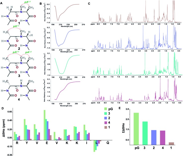
Fig. 3. (A) Interplay of pseudoallylic strain (pA1,2 and pA1,3) in the reverse turn to induce the β-sheet folding. Note the spatial positioning of the methyl groups between i+1 Cα and i+2 Cα which modulates the folding of the polypeptide in aqueous solution at 25 °C. (B) CD spectrum of the peptides in acetate buffer (pH 3.8). (C) 1H NMR in acetate buffer (pH 3.8), clearly indicating the enhanced dispersion of the HN resonance and upfield shifted, distinguishable Leu11 Hδ1/2 in the well-folded peptides. (D) Secondary chemical shifts obtained from the respective random coil control peptides, where the d-residue is substituted with the l-residue. Leu11 Hα shows a negative deviation due to its proximity to the phenyl ring of Tyr2 in the folded analogs. In 1, the secondary chemical shifts were determined by subtracting the Hα chemical shifts from the mean random coil chemical shifts of the respective residues obtained from the random coils of 2 and 3. The ring constrained reverse turn motif, pG, was taken as a reference.19 (E) Summation of the secondary chemical shifts; only the absolute values were considered.

Fig. 3