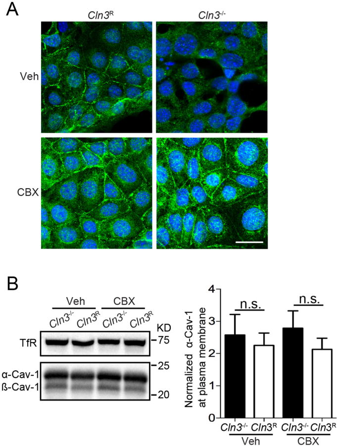
Fig. 4.

CBX restores Cav-1 subcellular localization in Cln3−/− MBECs. A, MBECs were treated with CBX (25 μM) or vehicle for 2 h, fixed, and Cav-1 intracellular distribution evaluated using immunocytochemistry and confocal microscopy. Photomicrographs are representative of 3 independent experiments. Scale bar = 20 μm.B, Western blotting of plasma membrane enriched fractions for Cav-1 levels and quantification. Data are from six independent experiments. Bars show mean ± s.e.m., No statistical differences were found by one-way ANOVA.
