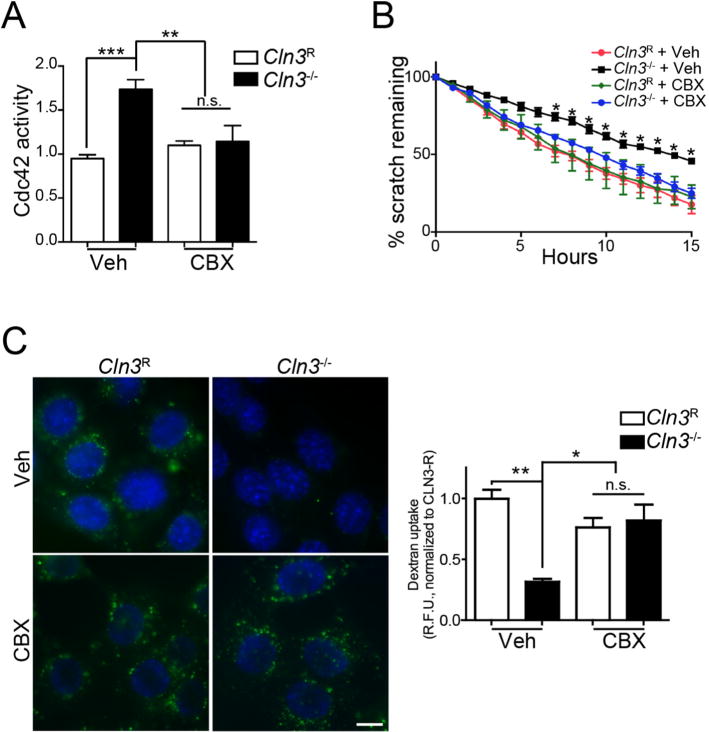
Fig. 5.

Correction of Cdc42-dependent defects in Cln3−/− MBECs with CBX. A, MBECs were treated for 2 h with CBX (50 μM) or vehicle and Cdc42-GTP levels were measured. Data are mean ± SEM of 5 independent experiments, one-way ANOVA with Tukey’s multiple comparison correction. B, MBECs were grown to confluence, a scratch wound made, and CBX (3.5 μM) or vehicle added to cells. Cell migration was imaged overnight and quantified by T-Scratch software. Data are mean ± s.e.m. from 4 independent experiments evaluated by one-way ANOVA with Tukey’s post-hoc correction. C, MBECs were treated with CBX (25 μM) or vehicle and endocytic uptake assessed by A488-Dextran uptake (green). Hoechst 33342 was used to label nuclei (blue). Non-internalized extracellular dextran was quenched by Red-40 and epifluorescent images were quantified by ImageJ. Data are mean ± s.e.m. from ~70 cells from 3 independent experiments, and evaluated one-way ANOVA with Tukey’s post-hoc. For all panels *, p < 0.05, **, p < 0.01, ***, p < 0.001, n.s. = not significant. (For interpretation of the references to colour in this figure legend, the reader is referred to the web version of this article.)
