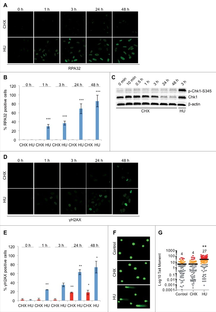
Figure 2.

CHX treatment does not result in ssDNA formation, checkpoint activation or DNA damage. (A) Representative images of U2OS cells treated with 10 μg/ml CHX or 2 mM HU for the indicated timepoints and stained for RPA32. Cells were pre-extracted with CSK buffer (10 mM Pipes, pH 7.0, 100 mM NaCl, 300 mM sucrose, and 3 mM MgCl2, 0.7% Triton X-100) for 5 minutes prior to fixation. (B) Quantification of RPA32 positive cells (A), (n = 3). (C) U2OS cells were either left untreated or treated with CHX or HU for the timepoints indicated followed by Western blot probed with p-Chk1-S345, Chk1 and β-actin antibodies. (D) Representative images of U2OS cells treated with 10 μg/ml CHX or 2 mM HU for the indicated timepoints and stained for γH2AX. (E) Quantification of γH2AX positive cells (D), (n = 2). (F) and (G) DNA damage in U2OS assessed by the comet assay following treatment with 10 μg/ml CHX or 2 mM HU for 24 hours. Representative images (F) and quantification of tail moment (G). The error bars depict standard deviation; *P ≤ 0.05, **P ≤ 0.01, ***P ≤ 0.001 as determined by Student's t-test. See also Figure S3.
