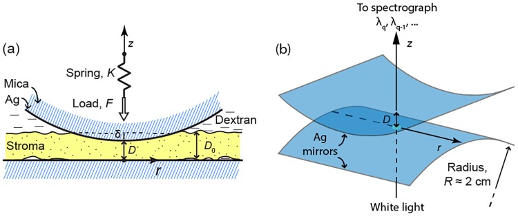
Fig 1. Schematic of the SFA setup for corneal compression and force measurements.
(A) Anterior corneal stroma with undeformed thickness D0 confined between the crossed cylindrical surfaces of the SFA. r is the lateral distance from the contact point, where the surface separation distance is D and thickness deformation is δ. The normal load F is applied via a double-cantilever spring with constant K whose fixed end undergoes step-wise vertical displacements (along z) as a function of time. (B) Curved Fabry-Perot optical interferometer used to determine D.

