
Fig. 3. RIPC and exogenous irisin protected against IR injury in the mouse lung.
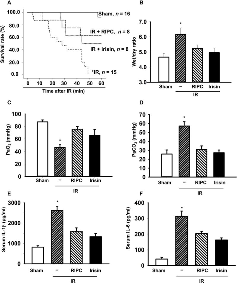
(A) The animal survival rate after lung IR injury with RIPC treatment or irisin administration (1 μg/kg) (*P < 0.05 versus others; n = 16 in sham group, n = 15 in IR group, and n = 8 in other groups). (B) Lung edema was evaluated as the wet/dry weight ratio of the excised lung from mice (*P < 0.05 versus others; n = 9 in sham group, n = 8 in IR group, and n = 4 in other groups). The plasma PaO2 (C) and PaCO2 (D) were measured (*P < 0.05 versus others; n = 8 in sham and IR group and n = 4 in other groups). A.U., arbitrary units. Serum concentrations of interleukin-1β (IL-1β) (E) and IL-6 (F) were measured using enzyme-linked immunosorbent assay (*P < 0.05 versus others; n = 4).